Search results
Found 12694 matches for
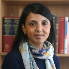
Dr Sushma Shankar from the Nuffield Department of Surgical Sciences (NDS) has been awarded the second prize for the very first European Society of Organ Transplant (ESOT) Leonard da Vinci Transplant Research Innovation Award, the most prestigious award of the ESOT 2019 Congress in Copenhagen.
Circulating TNFα in deceased donors promotes kidney injury and associates with inferior short- and long-term graft function and survival.
In deceased donation, donor management and organ procurement may contribute to donor organ injury, particularly through triggering systemic inflammation. Despite the important clinical implications, the impact of circulating inflammation on donor kidney injury, and short- and long-term posttransplant outcomes are unknown. We quantified TNFα and its receptors TNFR1 and TNFR2 in 1018 longitudinal plasma samples collected during donor management from 596 deceased and 34 living donors, from multiple centres across the United Kingdom. High donor plasma TNFα levels significantly associated with 12 and up to 60 months inferior graft function and up to 96 months reduced survival, only in DBDs but not in DCDs. Associations were replicated in a validation cohort and withstood linear mixed model adjustments for donor and recipient covariates. Analysis of paired plasma and kidney biopsy samples revealed that high plasma TNFα levels correlated with increased expression of injury markers in donor kidney. Further in vitro investigations confirmed that human podocytes, exposed to TNFα donor plasma, demonstrated TNFR1 signaling driven injury profiles, a response that was ameliorated by infliximab. Our data provide evidence that monitoring plasma inflammation levels during donor management offers a window of opportunity to intervene and improve optimisation and quality of deceased donor organs.
Adiponectin Reduces Glomerular Endothelial Glycocalyx Disruption and Restores Glomerular Barrier Function in a Mouse Model of Type 2 Diabetes
Adiponectin has vascular anti-inflammatory and protective effects. Although adiponectin protects against the development of albuminuria, historically, the focus has been on podocyte protection within the glomerular filtration barrier (GFB). The first barrier to albumin in the GFB is the endothelial glycocalyx (eGlx), a surface gel-like barrier covering glomerular endothelial cells (GEnCs). In diabetes, eGlx dysfunction occurs before podocyte damage; hence, we hypothesized that adiponectin could protect from eGlx damage to prevent early vascular damage in diabetic kidney disease (DKD). Globular adiponectin (gAd) activated AMPK signaling in human GEnCs through AdipoR1. It significantly reduced eGlx shedding and the tumor necrosis factor-α (TNF-α)–mediated increase in syndecan-4 (SDC4) and MMP2 mRNA expression in GEnCs in vitro. It protected against increased TNF-α mRNA expression in glomeruli isolated from db/db mice and against expression of genes associated with glycocalyx shedding (namely, SDC4, MMP2, and MMP9). In addition, gAd protected against increased glomerular albumin permeability (Ps’alb) in glomeruli isolated from db/db mice when administered intraperitoneally and when applied directly to glomeruli (ex vivo). Ps’alb was inversely correlated with eGlx depth in vivo. In summary, adiponectin restored eGlx depth, which was correlated with improved glomerular barrier function, in diabetes. Article Highlights
Heparanase inhibition as a systemic approach to protect the endothelial glycocalyx and prevent microvascular complications in diabetes
Abstract Background Diabetes mellitus is a chronic disease which is detrimental to cardiovascular health, often leading to secondary microvascular complications, with huge global health implications. Therapeutic interventions that can be applied to multiple vascular beds are urgently needed. Diabetic retinopathy (DR) and diabetic kidney disease (DKD) are characterised by early microvascular permeability changes which, if left untreated, lead to visual impairment and renal failure, respectively. The heparan sulphate cleaving enzyme, heparanase, has previously been shown to contribute to diabetic microvascular complications, but the common underlying mechanism which results in microvascular dysfunction in conditions such as DR and DKD has not been determined. Methods In this study, two mouse models of heparan sulphate depletion (enzymatic removal and genetic ablation by endothelial specific Exotosin-1 knock down) were utilized to investigate the impact of endothelial cell surface (i.e., endothelial glycocalyx) heparan sulphate loss on microvascular barrier function. Endothelial glycocalyx changes were measured using fluorescence microscopy or transmission electron microscopy. To measure the impact on barrier function, we used sodium fluorescein angiography in the eye and a glomerular albumin permeability assay in the kidney. A type 2 diabetic (T2D, db/db) mouse model was used to determine the therapeutic potential of preventing heparan sulphate damage using treatment with a novel heparanase inhibitor, OVZ/HS-1638. Endothelial glycocalyx changes were measured as above, and microvascular barrier function assessed by albumin extravasation in the eye and a glomerular permeability assay in the kidney. Results In both models of heparan sulphate depletion, endothelial glycocalyx depth was reduced and retinal solute flux and glomerular albumin permeability was increased. T2D mice treated with OVZ/HS-1638 had improved endothelial glycocalyx measurements compared to vehicle treated T2D mice and were simultaneously protected from microvascular permeability changes associated with DR and DKD. Conclusion We demonstrate that endothelial glycocalyx heparan sulphate plays a common mechanistic role in microvascular barrier function in the eye and kidney. Protecting the endothelial glycocalyx damage in diabetes, using the novel heparanase inhibitor OVZ/HS-1638, effectively prevents microvascular permeability changes associated with DR and DKD, demonstrating a novel systemic approach to address diabetic microvascular complications.
Reduced Glomerular Filtration in Diabetes Is Attributable to Loss of Density and Increased Resistance of Glomerular Endothelial Cell Fenestrations
Significance Statement We propose a novel mechanism underlying loss of renal filtration function from studying glomerular endothelial cell (GEnC) fenestrae in human diabetic kidney tissue and in a mouse model of diabetes. Diaphragmed fenestrae may provide structural resistance to filtration. We hypothesize that EHD3 is a key regulator of GEnC fenestrations, and its glomerular expression is lost in diabetes. This study establishes the critical role of GEnC fenestrations in renal filtration function and suggests a key regulator, potentially paving the way for development of targeted therapies to restore fenestrae and thus filtration function in kidney disease. Background Glomerular endothelial cell (GEnC) fenestrations are recognized as an essential component of the glomerular filtration barrier, yet little is known about how they are regulated and their role in disease. Methods We comprehensively characterized GEnC fenestral and functional renal filtration changes including measurement of glomerular K f and GFR in diabetic mice (BTBR ob−/ob− ). We also examined and compared human samples. We evaluated Eps homology domain protein-3 (EHD3) and its association with GEnC fenestrations in diabetes in disease samples and further explored its role as a potential regulator of fenestrations in an in vitro model of fenestration formation using b.End5 cells. Results Loss of GEnC fenestration density was associated with decreased filtration function in diabetic nephropathy. We identified increased diaphragmed fenestrations in diabetes, which are posited to increase resistance to filtration and further contribute to decreased GFR. We identified decreased glomerular EHD3 expression in diabetes, which was significantly correlated with decreased fenestration density. Reduced fenestrations in EHD3 knockdown b.End5 cells in vitro further suggested a mechanistic role for EHD3 in fenestration formation. Conclusions This study demonstrates the critical role of GEnC fenestrations in renal filtration function and suggests EHD3 may be a key regulator, loss of which may contribute to declining glomerular filtration function through aberrant GEnC fenestration regulation. This points to EHD3 as a novel therapeutic target to restore filtration function in disease.
Endothelial glycocalyx is damaged in diabetic cardiomyopathy: angiopoietin 1 restores glycocalyx and improves diastolic function in mice
Abstract Aims/hypothesis Diabetic cardiomyopathy (DCM) is a serious and under-recognised complication of diabetes. The first sign is diastolic dysfunction, which progresses to heart failure. The pathophysiology of DCM is incompletely understood but microcirculatory changes are important. Endothelial glycocalyx (eGlx) plays multiple vital roles in the microcirculation, including in the regulation of vascular permeability, and is compromised in diabetes but has not previously been studied in the coronary microcirculation in diabetes. We hypothesised that eGlx damage in the coronary microcirculation contributes to increased microvascular permeability and hence to cardiac dysfunction. Methods We investigated eGlx damage and cardiomyopathy in mouse models of type 1 (streptozotocin-induced) and type 2 (db/db) diabetes. Cardiac dysfunction was determined by echocardiography. We obtained eGlx depth and coverage by transmission electron microscopy (TEM) on mouse hearts perfusion-fixed with glutaraldehyde and Alcian Blue. Perivascular oedema was assessed from TEM images by measuring the perivascular space area. Lectin-based fluorescence was developed to study eGlx in paraformaldehyde-fixed mouse and human tissues. The eGlx of human conditionally immortalised coronary microvascular endothelial cells (CMVECs) in culture was removed with eGlx-degrading enzymes before measurement of protein passage across the cell monolayer. The mechanism of eGlx damage in the diabetic heart was investigated by quantitative reverse transcription-PCR array and matrix metalloproteinase (MMP) activity assay. To directly demonstrate that eGlx damage disturbs cardiac function, isolated rat hearts were treated with enzymes in a Langendorff preparation. Angiopoietin 1 (Ang1) is known to restore eGlx and so was used to investigate whether eGlx restoration reverses diastolic dysfunction in mice with type 1 diabetes. Results In a mouse model of type 1 diabetes, diastolic dysfunction (confirmed by echocardiography) was associated with loss of eGlx from CMVECs and the development of perivascular oedema, suggesting increased microvascular permeability. We confirmed in vitro that eGlx removal increases CMVEC monolayer permeability. We identified increased MMP activity as a potential mechanism of eGlx damage and we observed loss of syndecan 4 consistent with MMP activity. In a mouse model of type 2 diabetes we found a similar loss of eGlx preceding the development of diastolic dysfunction. We used isolated rat hearts to demonstrate that eGlx damage (induced by enzymes) is sufficient to disturb cardiac function. Ang1 restored eGlx and this was associated with reduced perivascular oedema and amelioration of the diastolic dysfunction seen in mice with type 1 diabetes. Conclusions/interpretation The association of CMVEC glycocalyx damage with diastolic dysfunction in two diabetes models suggests that it may play a pathophysiological role and the enzyme studies confirm that eGlx damage is sufficient to impair cardiac function. Ang1 rapidly restores the CMVEC glycocalyx and improves diastolic function. Our work identifies CMVEC glycocalyx damage as a potential contributor to the development of DCM and therefore as a therapeutic target. Graphical abstract
Past, Present, and Future of Dynamic Kidney and Liver Preservation and Resuscitation.
The increased demand for organs has led to the increased usage of "higher risk" kidney and liver grafts. These grafts from donation after circulatory death or expanded criteria donors are more susceptible to preservation injury and have a higher risk of unfavorable outcomes. Dynamic, instead of static, preservation could allow for organ optimization, offering a platform for viability assessment, active organ repair and resuscitation. Ex situ machine perfusion and in situ regional perfusion in the donor are emerging as potential tools to preserve and resuscitate vulnerable grafts. Preclinical findings have ignited clinical organ preservation research that investigates dynamic preservation, its various modes (continuous, preimplantation) and temperatures (hypo-, sub, or normothermic). This review outlines the current status of dynamic preservation of kidney and liver grafts and describes ongoing research and emerging clinical trials.
Using an Integrated -Omics Approach to Identify Key Cellular Processes That Are Disturbed in the Kidney After Brain Death.
In an era where we are becoming more reliant on vulnerable kidneys for transplantation from older donors, there is an urgent need to understand how brain death leads to kidney dysfunction and, hence, how this can be prevented. Using a rodent model of hemorrhagic stroke and next-generation proteomic and metabolomic technologies, we aimed to delineate which key cellular processes are perturbed in the kidney after brain death. Pathway analysis of the proteomic signature of kidneys from brain-dead donors revealed large-scale changes in mitochondrial proteins that were associated with altered mitochondrial activity and morphological evidence of mitochondrial injury. We identified an increase in a number of glycolytic proteins and lactate production, suggesting a shift toward anaerobic metabolism. Higher amounts of succinate were found in the brain death group, in conjunction with increased markers of oxidative stress. We characterized the responsiveness of hypoxia inducible factors and found this correlated with post-brain death mean arterial pressures. Brain death leads to metabolic disturbances in the kidney and alterations in mitochondrial function and reactive oxygen species generation. This metabolic disturbance and alteration in mitochondrial function may lead to further cellular injury. Conditioning the brain-dead organ donor by altering metabolism could be a novel approach to ameliorate this brain death-induced kidney injury.
Plasma degradome affected by variable storage of human blood.
BACKGROUND: The successful application of-omics technologies in the discovery of novel biomarkers and targets of therapeutic interventions is facilitated by large collections of well curated clinical samples stored in bio banks. Mining the plasma proteome holds promise to improve our understanding of disease mechanisms and may represent a source of biomarkers. However, a major confounding factor for defining disease-specific proteomic signatures in plasma is the variation in handling and processing of clinical samples leading to protein degradation. To address this, we defined a plasma proteolytic signature (degradome) reflecting pre-analytical variability in blood samples that remained at ambient temperature for different time periods after collection and prior to processing. METHODS: We obtained EDTA blood samples from five healthy volunteers (n = 5), and blood tubes remained at ambient temperature for 30 min, 8, 24 and 48 h prior to centrifugation and isolation of plasma. Naturally occurred peptides derived from plasma samples were compared by label-free quantitative LC-MS/MS. To profile protein degradation, we analysed pooled plasma samples at T = 30 min and 48 h using PROTOMAP analysis. The proteolytic pattern of selected protein candidates was further validated by immunoblotting. RESULTS: A total of 820 plasma proteins were surveyed by PROTOMAP, and for 4 % of these, marked degradation was observed. We show distinct proteolysis patterns for talin-1, coagulation factor XI, complement protein C1r, C3, C4 and thrombospondin, and several proteins including S100A8, A9, annexin A1, profiling-1 and platelet glycoprotein V are enriched after 48 h blood storage at ambient temperature. In particular, thrombospondin protein levels increased after 8 h and proteolytic fragments appeared after 24 h storage time. CONCLUSIONS: The overall impact of blood storage at ambient temperature for variable times on the plasma proteome and degradome is relatively minor, but in some cases can cause a potential bias in identifying and assigning relevant proteomic markers. The observed effects on the plasma proteome and degradome are predominantly triggered by limited leucocyte and platelet cell activation due to blood handling and storage. The baseline plasma degradome signature presented here can help filtering candidate protein markers relevant for clinical biomarker studies.
Oxygenated versus standard cold perfusion preservation in kidney transplantation (COMPARE): a randomised, double-blind, paired, phase 3 trial.
BACKGROUND: Deceased donor kidneys are preserved in cold hypoxic conditions. Providing oxygen during preservation might improve post-transplant outcomes, particularly for kidneys subjected to greater degrees of preservation injury. This study aimed to investigate whether supplemental oxygen during hypothermic machine perfusion (HMP) could improve the outcome of kidneys donated after circulatory death. METHODS: This randomised, double-blind, paired, phase 3 trial was done in 19 European transplant centres. Kidney pairs from donors aged 50 years or older, donated after circulatory death, were eligible if both kidneys were transplanted into two different recipients. One kidney from each donor was randomly assigned using permuted blocks to oxygenated hypothermic machine perfusion (HMPO2), the other to HMP without oxygenation. Perfusion was maintained from organ retrieval to implantation. The primary outcome was 12-month estimated glomerular filtration rate (eGFR) using the Chronic Kidney Disease Epidemiology Collaboration equation in pairs of donated kidneys in which both transplanted kidneys were functioning at the end of follow-up. Safety outcomes were reported for all transplanted kidneys. Intention-to-treat analyses were done. This trial is registered with the ISRCTN Registry, ISRCTN32967929, and is now closed. FINDINGS: Between March 15, 2015, and April 11, 2017, 197 kidney pairs were randomised with 106 pairs transplanted into eligible recipients. 23 kidney pairs were excluded from the primary analysis because of kidney failure or patient death. Mean eGFR at 12 months was 50·5 mL/min per 1·73 m2 (SD 19·3) in the HMPO2 group versus 46·7 mL/min per 1·73m2 (17·1) in HMP (mean difference 3·7 mL/min per 1·73m2, 95% CI -1·0 to 8·4; p=0·12). Fewer severe complications (Clavien-Dindo grade IIIb or more) were reported in the HMPO2 group (46 of 417, 11%, 95% CI 8% to 14%) than in the HMP group (76 of 474, 16%, 13% to 20%; p=0·032). Graft failure was lower with HMPO2 (three [3%] of 106) compared with HMP (11 [10%] of 106; hazard ratio 0·27, 95% CI 0·07 to 0·95; p=0·028). INTERPRETATION: HMPO2 of kidneys donated after circulatory death is safe and reduces post-transplant complications (grade IIIb or more). The 12-month difference in eGFR between the HMPO2 and HMP groups was not significant when both kidneys from the same donor were still functioning 1-year post-transplant, but potential beneficial effects of HMPO2 were suggested by analysis of secondary outcomes. FUNDING: European Commission 7th Framework Programme.
Cytoskeletal protein degradation in brain death donor kidneys associates with adverse posttransplant outcomes.
In brain death, cerebral injury contributes to systemic biological dysregulation, causing significant cellular stress in donor kidneys adversely impacting the quality of grafts. Here, we hypothesized that donation after brain death (DBD) kidneys undergo proteolytic processes that may deem grafts susceptible to posttransplant dysfunction. Using mass spectrometry and immunoblotting, we mapped degradation profiles of cytoskeletal proteins in deceased and living donor kidney biopsies. We found that key cytoskeletal proteins in DBD kidneys were proteolytically cleaved, generating peptide fragments, predominantly in grafts with suboptimal posttransplant function. Interestingly, α-actinin-4 and talin-1 proteolytic fragments were detected in brain death but not in circulatory death or living donor kidneys with similar donor characteristics. As talin-1 is a specific proteolytic target of calpain-1, we investigated a potential trigger of calpain activation and talin-1 degradation using human ex vivo precision-cut kidney slices and in vitro podocytes. Notably, we showed that activation of calpain-1 by transforming growth factor-β generated proteolytic fragments of talin-1 that matched the degradation fragments detected in DBD preimplantation kidneys, also causing dysregulation of the actin cytoskeleton in human podocytes; events that were reversed by calpain-1 inhibition. Our data provide initial evidence that brain death donor kidneys are more susceptible to cytoskeletal protein degradation. Correlation to posttransplant outcomes may be established by future studies.
Renal biopsies from donors with acute kidney injury show different molecular patterns according to the post-transplant function.
The utilization of kidneys from donors with acute kidney injury (AKI) is often limited by unpredictable post-transplantation outcomes. The aim of our study was to identify protein mediators implicated in either recovery or failure of these organs. Forty kidney biopsies from donors with (20) and without AKI (20) were selected and then subdivided according to the post-transplant outcome defined as a threshold of 45 ml/min for the eGFR at 1 year from transplantation. Tissue homogenates were analysed by western blot to assess how the levels of 17 pre-selected proteins varied across the four groups. Samples from AKI kidneys with a poor outcome showed a fourfold increase in the levels of PPARg and twofold reduction of STAT1 compared to the other groups (p