Multidisciplinary Research Group
Novel Applications in Multimodal and Digital Pathology
includes Oxford Biocore - The NDS Tissue Handling Platform
We aim to embed the role of pathology across all research for the benefit of patients and to bring about improvements in the quality of reseach. Our team have interests in a wide range of projects including in digital pathology and image analysis, and multimodal and molecular pathology. Our group also includes Oxford Biocore, the recently formed NDS Tissue Handling Platform.

Histopathology, the microscopic study of diseased tissue, is an important tool in research, since accurate diagnosis and analysis of cancer and other diseases allows researchers to better understand the effects on patients and their prognosis. It enables clinicians to stratify patients into risk groups and, in some cases, can ensure patients are not over or under treated. Samples of tissue are often stained for various markers to help diagnose and understand abnormal cells found in tumours, and to detect cellular events such as cell death which may help in understanding the behaviours in certain cancers, and the implications for disease progression. For instance, using immunohistochemistry staining for expression of a protein kinase in prostate cancer may be a marker of response to radiotherapy allowing clinicians to determine the effectiveness of the treatment.
Molecular pathology assesses the changes in tissues together with an understanding of the molecular and genetic changes in human diseases (especially cancer), and includes the design and validation of predictive biomarkers for treatment response and disease progression. It enables researchers to understand why individuals of different genetic constitutions go on to develop disorders whereas others do not. Various techniques are used in molecular pathology and include quantitative polymerase chain reaction (qPCR), multiplex PCR, DNA microarray and DNA sequencing, and antibody based immunofluorescence tissue assays. More accurate diagnosis is possible when the diagnosis is based on both the morphologic changes in tissues and on molecular testing. For example, the 100,000 Genomes Project aims to sequence the DNA from around 70,000 patients. By comparing DNA from a patient’s tumour and healthy cells researchers will gain insight into the exact nature and genomic changes that are causing an individual’s cancer. For more information, see: https://www.genomicsengland.co.uk/the-100000-genomes-project/#
Multimodal pathology and digital pathology enables researchers to convert glass slides, which have usually been stained with various markers, to digitised images and to use various tools such as machine learning programmes to determine various histological features. Algorithms can be used to automate processes such as the manual counting of cell types, or to determine the number and type of cells in tumour and non tumour tissue. The aim is to reduce human error, improve accuracy of diagnosis and enable researchers to determine the differences in cell type, number and architecture in various cancers and other diseases and the effect on patient outcome. For instance, by using an algorithm to assess the different density/distribution of tumour infiltrating lymphocytes in both cancer and non cancer tissue, and using this information in association with information about the patient’s disease progression,e.g the clinical stage at presentation or disease relapse , it may be possible to improve patient prognosis in the longer term by determining histological features in testicular tumours and their association with outcome.
Digital slides are also easier to share than physical slides and could lead to developments allowing better patient outcomes such as enabling experts to consult on rare cases or to take part in virtual multi-disciplinary team meetings (MDT’s) where consultations can take place much more quickly and efficiently.

Members of the Verrill Pathology Group
Our team
-
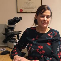
Clare Verrill
Professor of Cellular Pathology and Artificial Intelligence
-
Monica Dolton
Research Project Manager / NDS Information Governance Manager
-
Adam Lambert
Bio-repository Manager
-
Lucia Cerundolo
Researcher
-
David Maldonado-Perez
Oxford Radcliffe Biobank (ORB) Collections Governance and Quality Manager
NEWS
Digital Pathology in Diagnostic Practice and AI - 7th & 8th January 2020, Oxford
PathLAKE and Oxford BRC are pleased to announce that they will be hosting a two day educational meeting to share the latest developments on implementing digital pathology into diagnostic practice in histopathology, together with an overview of current and future applications of digital pathology in the research and AI setting. Guest speakers will be sharing real life experience, discussing research applications and will provide an overview of best practice throughout the two days.
The meeting is aimed at diagnostic histopathologists (trainees and consultants) and may also be of interest to biomedical scientists.
Register by emailing denise.bent@philips.com. The event is free to attend but spaces are limited, and will be offered on a first-come first-served basis.
Also see the PathLAKE website: click here

Professor Verrill leads The Oxford Centre for PathLAKE with £2.7m award
The Oxford centre for Project PathLAKE has been awarded £2.7m from government and industry, funded through the Industrial Strategy Challenge Fund, the government’s flagship investment programme, which focuses on addressing the opportunities and challenges of the future, and which is managed by UK Research and Innovation.
Oxford PathLAKE is led by Dr Clare Verrill, from NDS, who also works in the OUH Cellular Pathology team, and who, with funding through the Oxford BRC’s Molecular Diagnostics Theme, was able to establish a successful programme on digital pathology and AI, which generated the pilot data that underpinned the application and allowed the team to take part in a collaborative programme proposal with Philips.
Oxford is a partner in the centres focusing on digital pathology, led by University Hospitals Coventry and Warwickshire NHS Trust. Healthcare technology company Philips will be the principal partner on this major cross-sector collaboration, called project PathLAKE (Pathology image data Lake for Education, Analytics and Discovery).
This initiative has received £10m of government funding in total to develop innovative AI in pathology. Project PathLAKE partners will embed and demonstrate the diagnostic efficiency of digital pathology, computer-aided testing of pathology samples, and develop novel AI tools to personalised medicine by selecting the right patients for the best therapy.
The project will create a secure data-lake of tens of thousands of professionally annotated images for building deep learning algorithms that can automatically detect cancer. These images and tools will be made available across the consortium including a growing number of SME partners in this sector to develop AI to overcome burgeoning workloads in the UK and establish a world-leading UK digital health industry.
Professor Verrill says:
"This is a tremendously exciting opportunity for Oxford to build on our programme in computational pathology, which is a novel collaboration between pathology (via NDS and the NHS Cellular Pathology lab at OUHFT) and Jens Rittscher, Professor of Engineering Science of the Big Data Institute. We will be changing the way cellular pathology is delivered in Oxford and across the UK, with deployment of novel digital reporting of live diagnostic cases and building and testing novel AI algorithms into the clinical workflow. We are incredibly proud to be a part of PathLAKE, a digital pathology consortium led by University Hospitals Coventry / The University of Warwick and backed by our industry partner Philips. This is a team effort and we would not have achieved this success without the input of several other key individuals, to name a few Kieron White and the NHS Cellular Pathology team and the Verrill and Rittscher research groups".
Professor Clare Verrill has played a major part in establishing a partnership between Oxford University Hospitals (OUH) NHS Foundation Trust and healthtech company Philips to create a digital pathology network to help drive faster and more efficient diagnoses for patients.
OUH will deploy Philips’ IntelliSite Pathology Solution to serve as a central laboratory for partner sites at Milton Keynes University Hospital NHS Foundation Trust and Great Western Hospital NHS Foundation Trust in Swindon.
Pathologists play a critical role in disease detection, particularly with cancer diagnosis. Traditionally, pathologists analyse tissue samples on glass slides under a microscope. When a pathologist requires a second opinion from a sub-specialist, these glass slides must be transported to a second site, which can result in lost or damaged slides or delays in diagnosis.
A digital network for pathology will allow clinicians across the three regions and within the Thames Valley Cancer Network to collaborate remotely on patient cases. It's hoped that remote collaboration will help reduce delays in slide transport times, encourage more efficient workflows across the sites, and enable quicker access to specialist pathology.
Professor Verrill said: "As an NHS Global Digital Exemplar committed to improving patient care by embracing the latest digital technologies and cross-site collaborations, this partnership aims to modernise patient care and offer innovative world-leading services".
The partnership has featured in several journals (see links below)
Clinical Services Journal
Building Better Healthcare
Digital Health Age
http://digitalhealthage.com/digital-pathology-network-to-speed-up-disease-diagnosis-in-oxford/
Health Investor
http://www.healthinvestor.co.uk/AccessDenied.aspx?ReturnUrl=/ShowArticle.aspx?ID=9638
Hospital Management
Compelo
https://www.compelo.com/medical-devices/news/philips-ouh-partner-digital-pathology-network/
BioPortfolio
Professor Verrill speaks at Innovation Forum 2017 - Leaders conference
Professor Verrill took part in the Innovation Forum meeting in December, speaking at the "Digital Health" session. The meeting was held in the Examination Schools in Oxford and brings together a mixture of leaders in industry, academia and the Government, together with investors and researchers.
Digital Pathology Congress - Global ENGAGE
Several members of the group attended this years 4th Digital Pathology Congress run by Global Engage in London. The event was entitled "UNDERSTANDING & UTILIZING DIGITAL PATHOLOGY AS A TOOL FOR ADVANCING PATHOLOGY PRACTICE & ENABLING ENHANCED PATIENT CARE"
Congratulations to Elysia Upton and Nasullah Khalid Alham who both presented posters. Professor Verrill and Dr Robert Pell gave a joint presentation at the meeting.
https://twitter.com/NDSurgicalSci/status/936527953235718145

from left to right: Dr Robert Pell, Elysia Upton and Dr Nasullah Khalid Alham at Global Engage
Professor Verrill undertakes key role within the National Cancer Research Institute
Professor Clare Verrill has recently been appointed the lead of workstream 4 (Technology and Informatics) within the Clinical Molecular Pathology (CM-Path) initiative.
To read more go to: http://www.ncri.org.uk/60-second-interviews/60-seconds-with-dr-clare-verrill/
Pathologists feature in videos for 100,000 Genomes Project
Professor Clare Verrill and Dr Lisa Browning have been filmed demonstrating how to sample fresh tumours from prostate and renal cases for whole genome sequencing and the 100,000 Genomes Project.
To read more go to: https://www.nds.ox.ac.uk/news/pathologists-feature-in-videos-for-100000-genomes-project
The biobank team feature in 100,000 Genomes Project Films
Members of the group, including Clare Verrill, Adam Lambert and Martin Taylor have taken part in a series of short educational films about sample processing for whole genome sequencing for the 100,000 Genomes Project.
To see the films use the link below:
http://www.nds.ox.ac.uk/news/biobank-team-feature-in-100-000-genomes-project-films
Giant touchscreen helping in the battle against cancer
Cancer Research UK Oxford Centre has funded the 55-inch wide screen for the teaching of histopathology speciality trainees and medical students from the University of Oxford and the 500 researchers and clinicians that make up the Oxford Centre
Clare Verrill taught the first session using the new screen and said: 'Having this digital screen, enables a new and exciting way of teaching where learners and teachers are able to interact with the images and makes for stimulating and interesting discussions.
'With an increasing trend for use of digital pathology images for routine diagnostics as well as research, histopathology registrars will be better equipped for modern pathology practice.'
For more information, see the link below:
http://www.nds.ox.ac.uk/news/giant-touchscreen-helping-in-the-battle-against-cancer
Collaborators
- Professor Johan Lundin and Dr Nina Lindar - Institute for Molecular Medicine Finland
https://www.fimm.fi/en/research/groups/lundin
- Professor Johann de Bono - Institute of Cancer Research, London
https://www.icr.ac.uk/our-research/researchers-and-teams/professor-johann-de-bono
- Professor Jens Rittscher - Institute of Biomedical Engineering, University of Oxford
http://www.ibme.ox.ac.uk/research/biomedia/jens-rittscher
- Dr Clare Turnbull - Institute of Cancer Research, London
https://www.icr.ac.uk/our-research/researchers-and-teams/dr-clare-turnbull
- Dr Srinivasa Rao Rao - Nuffield Dept of Surgical Sciences, University of Oxford
https://www.researchgate.net/profile/Srinivasa_Rao_Rao
- Professor Freddie Hamdy - Nuffield Dept of Surgical Sciences, University of Oxford
https://www.nds.ox.ac.uk/team/freddie-hamdy
- Professor Andrew Protheroe - Department of Oncology, University of Oxford
- Professor Gareth Bond - Nuffield Dept of Medicine, University of Oxford
https://www.ndm.ox.ac.uk/principal-investigators/researcher/gareth-bond
- Professor Adrian Hill - Nuffield Dept of Medicine, University of Oxford
https://www.ndm.ox.ac.uk/principal-investigators/researcher/adrian-hill
- Dr Prasanna Sooriakumaran - Nuffield Dept of Surgical Sciences, University of Oxford
https://www.nds.ox.ac.uk/team/prasanna-sooriakumaran
- Dr David Wedge - Nuffield Dept of Medicine, University of Oxford
https://www.ndm.ox.ac.uk/principal-investigators/researcher/david-wedge
- Dr Claire Palles - Nuffield Dept of Medicine, University of Oxford
https://www.ndm.ox.ac.uk/principal-investigators/researcher/claire-palles